Tab. II · Interactive anatomy
Surgical Anatomy Models
3D models of organs and tissues commonly encountered in general and gastrointestinal surgery. Choose an organ, then drag to rotate the model or use the preset angle buttons. The location diagram and text explain where each organ sits and why it matters in surgery.
Organs · 6
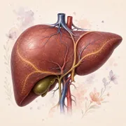
The gallbladder model is derived from an anatomical database (BodyParts3D), and its structure names come from that source. The other five are AI-generated artistic models: their surface vessels, colours and orientation do not reproduce real anatomy. Rely on the text and the location diagram for accurate positions and relations.
Drag to rotate and use the mouse wheel to zoom. With the model focused, the arrow keys rotate it and Page Up / Page Down zoom.Swipe sideways to rotate and pinch to zoom, or use the buttons.
01 · Hepar
Liver
The liver sits in the upper right abdomen. It makes bile and helps process nutrients.
Surgical relevance
In liver surgery, the relationship of each segment to its vessels and bile ducts matters. The surface vessels on this model are artistic and do not show the real vascular pattern; in reality the branches of the portal vein, hepatic artery and hepatic veins run inside the liver tissue. Couinaud segments are not separated in the model.
Detailed anatomy
Structure and parts
The liver lies below the diaphragm. Right and left lobes are visible externally; vascular and biliary anatomy defines eight functional segments.
Blood supply and relations
The portal vein and hepatic artery supply blood. The porta hepatis carries these vessels and bile ducts; hepatic veins drain to the inferior vena cava.
Common reasons for assessment
Anatomy matters when assessing cysts, masses and liver dysfunction.
- System
- Hepatobiliary system
- Model
- AI-generated artistic model; a single unnamed surface. Vessels, colours and orientation are unverified.
- Markers
- None; structures are described in the text
- File
- glTF · 2.7 MB
- Source
- NCBI Bookshelf: Gross and Surgical Anatomy of the Liver
In the digestive tract atlas
Related conditions, diagnostic and treatment methods, and research in this area.
02 · Vesica biliaris
Gallbladder
The gallbladder is a small organ beneath the liver that stores bile.
Surgical relevance
During cholecystectomy, the cystic duct and cystic artery must be identified safely before they are divided; the critical view of safety (CVS) approach is used for this. Anatomical variation must be considered.
The three criteria of the critical view of safety (SAGES)
- The hepatocystic triangle (bounded by the cystic duct, the common hepatic duct and the inferior edge of the liver) is cleared of fat and fibrous tissue.
- The lower third of the gallbladder is separated from the liver to expose the cystic plate.
- Two and only two structures, the cystic duct and the cystic artery, are seen entering the gallbladder.
Structures in the model
Numbers match the markers on the model; names and positions come from the source model.
- Fundus
- Body
- Infundibulum
- Neck
- Bile ducts
Detailed anatomy
Structure and parts
It lies on the liver's lower surface near segments IV and V. It has a fundus, body, infundibulum and neck; the neck continues into the cystic duct.
Blood supply and relations
The cystic duct joins the common hepatic duct to form the common bile duct. The cystic artery has variable anatomy.
Common reasons for assessment
This anatomy is relevant to gallstones and gallbladder inflammation.
- System
- Hepatobiliary system
- Model
- Derived from an anatomical database (BodyParts3D / Z-Anatomy); gallbladder and bile ducts are separate named objects
- Markers
- 5 structures; names and positions from the source model
- File
- glTF · 63 kB
- Source
- NCBI Bookshelf: Gallbladder Anatomy · SAGES: Safe Cholecystectomy Multi-Society Practice Guideline
In the digestive tract atlas
Related conditions, diagnostic and treatment methods, and research in this area.
03 · Pancreas
Pancreas
The pancreas lies behind the stomach and produces digestive secretions and hormones that regulate blood sugar.
Surgical relevance
Surgery near the head requires joint assessment of the duodenum, the common bile duct and the major vessels.
Detailed anatomy
Structure and parts
The head, neck, body, tail and uncinate process are described. The head sits in the duodenal C-loop; the main pancreatic duct drains into the duodenum.
Blood supply and relations
The head and duodenum share a pancreaticoduodenal vascular network. The neck lies in front of the point where the superior mesenteric and splenic veins join to form the portal vein.
Common reasons for assessment
These relationships matter in pancreatitis, cysts and masses.
- System
- Digestive and endocrine system
- Model
- AI-generated artistic model; a single unnamed surface. Vessels, colours and orientation are unverified.
- Markers
- None; structures are described in the text
- File
- glTF · 4.8 MB
- Source
- NCBI Bookshelf: Pancreas Anatomy
In the digestive tract atlas
Related conditions, diagnostic and treatment methods, and research in this area.
04 · Intestinum tenue et crassum
Intestines
The small intestine absorbs nutrients; the large intestine helps recover water.
Surgical relevance
Bowel resection requires attention to blood supply, adjacent organs and a tension-free anastomosis (the join between two bowel ends).
Detailed anatomy
Structure and parts
The small intestine comprises duodenum, jejunum and ileum. The large intestine includes caecum, appendix, ascending, transverse, descending and sigmoid colon, and rectum.
Blood supply and relations
Most of the small bowel and the right colon are supplied by the superior mesenteric artery (SMA); the left colon by the inferior mesenteric artery (IMA). The two territories are linked by the marginal artery running along the colon; at junction (watershed) zones such as the splenic flexure, the blood supply can be more tenuous.
Common reasons for assessment
Segmental anatomy helps assess appendicitis, diverticular disease, obstruction and colorectal disease.
- System
- Digestive system
- Model
- AI-generated artistic model; a single unnamed surface. Vessels, colours and orientation are unverified.
- Markers
- None; structures are described in the text
- File
- glTF · 2.0 MB
- Source
- NCBI Bookshelf: Large Intestine · Small Bowel
In the digestive tract atlas
Related conditions, diagnostic and treatment methods, and research in this area.
05 · Renes
Kidneys
The kidneys lie on either side of the spine and filter waste into urine.
Surgical relevance
In retroperitoneal and colonic surgery, knowing where the ureter and renal vessels lie matters. The vessel bridge joining the two kidneys in the model is an artistic simplification: in reality each renal artery arises separately from the aorta, each renal vein drains separately into the inferior vena cava, and at the hilum the usual front-to-back order is vein, artery, renal pelvis. Internal regions are not visible in this surface model.
Detailed anatomy
Structure and parts
They lie on the posterior abdominal wall, behind the peritoneum (retroperitoneal). The outer cortex, inner medulla and urine-collecting renal pelvis are key regions.
Blood supply and relations
The renal vessels pass through the hilum on the kidney's inner border. The pelvis continues as the ureter. The liver and duodenum lie near the right kidney; the spleen and pancreatic tail near the left.
Common reasons for assessment
Cross-sectional anatomy is used when assessing stones, cysts and masses.
- System
- Urinary system
- Model
- AI-generated artistic model; a single unnamed surface. Vessels, colours and orientation are unverified.
- Markers
- None; structures are described in the text
- File
- glTF · 2.2 MB
- Source
- NCBI Bookshelf: Kidneys
06 · Cutis et tela subcutanea
Skin & Soft Tissue
Skin protects the body. The soft tissue below includes fat and connective tissue.
Surgical relevance
Layer depth matters when assessing superficial lesions and planning incisions. In abdominal surgery the incision passes through skin, subcutaneous fat and the musculoaponeurotic layers of the abdominal wall to reach the peritoneum.
Detailed anatomy
Structure and parts
The epidermis is outermost, with dermis beneath and subcutaneous fat deeper still. Hair follicles and sweat and sebaceous glands lie in the dermis.
Blood supply and relations
The dermis and subcutaneous tissue contain vascular networks. Thickness varies by body region.
Common reasons for assessment
Layer anatomy is useful when assessing lipomas, skin cysts and wounds.
- System
- Integumentary system
- Model
- AI-generated artistic model; a single unnamed surface. Vessels, colours and orientation are unverified.
- Markers
- None; structures are described in the text
- File
- glTF · 5.8 MB
- Source
- NCBI Bookshelf: Skin
In the digestive tract atlas
Related conditions, diagnostic and treatment methods, and research in this area.
Tab. I · Digestive tract atlas
The models, diagrams and descriptions on this page are for general information only; models and diagrams are not to scale and do not replace individual diagnosis or treatment advice.
Sources last checked: 24 September 2026. Physician review details have not yet been published.
The gallbladder model is adapted from the Z-Anatomy atlas (originally BodyParts3D, The Database Center for Life Science) and is shared under the same CC BY-SA 4.0 licence.
The other five models and the thumbnails come from the thebuggeddev/anatomy model set; they are AI-generated (Tripo) artistic models.